Search Thermo Fisher Scientific
Disclaimer

Clicking the images or links will redirect you to a website hosted by BenchSci that provides third-party scientific content. Neither the content nor the BenchSci technology and processes for selection have been evaluated by us; we are providing them as-is and without warranty of any kind, including for use or application of the Thermo Fisher Scientific products presented.
Invitrogen
CD146 Monoclonal Antibody (P1H12), NovaFluor™ Blue 660-120S, eBioscience™
{{$productOrderCtrl.translations['antibody.pdp.commerceCard.promotion.promotions']}}
{{$productOrderCtrl.translations['antibody.pdp.commerceCard.promotion.viewpromo']}}
{{$productOrderCtrl.translations['antibody.pdp.commerceCard.promotion.promocode']}}: {{promo.promoCode}} {{promo.promoTitle}} {{promo.promoDescription}}. {{$productOrderCtrl.translations['antibody.pdp.commerceCard.promotion.learnmore']}}
")
FIGURE: 1 / 2
CD146 Antibody (H058T03B08-A) in Flow
Normal human peripheral blood cells were unstained (left) or stained with CD146 Monoclonal Antibody, NovaFluor Blue 660-120S (Product #: h058t03b08) (right). All cells were co-stained with CD3 Monoclonal Antibody, eFluor 450 (Product #: 48-0037-42). Total viable cells in the lymphocyte gate were used for analysis, as determined by LIVE/DEAD Blue (Product # L34962). Data was acquired on a 5-laser Cytek Aurora and unmixed with autofluorescence extraction.


 in Flow")
 in Flow")
Product Details
H058T03B08-A
Product Specifications
Species Reactivity
Dog,
Human,
Mouse,
Rabbit
Host/Isotype
Mouse
/ IgG1, kappa
Class
Monoclonal
Type
Antibody
Clone
P1H12
Conjugate
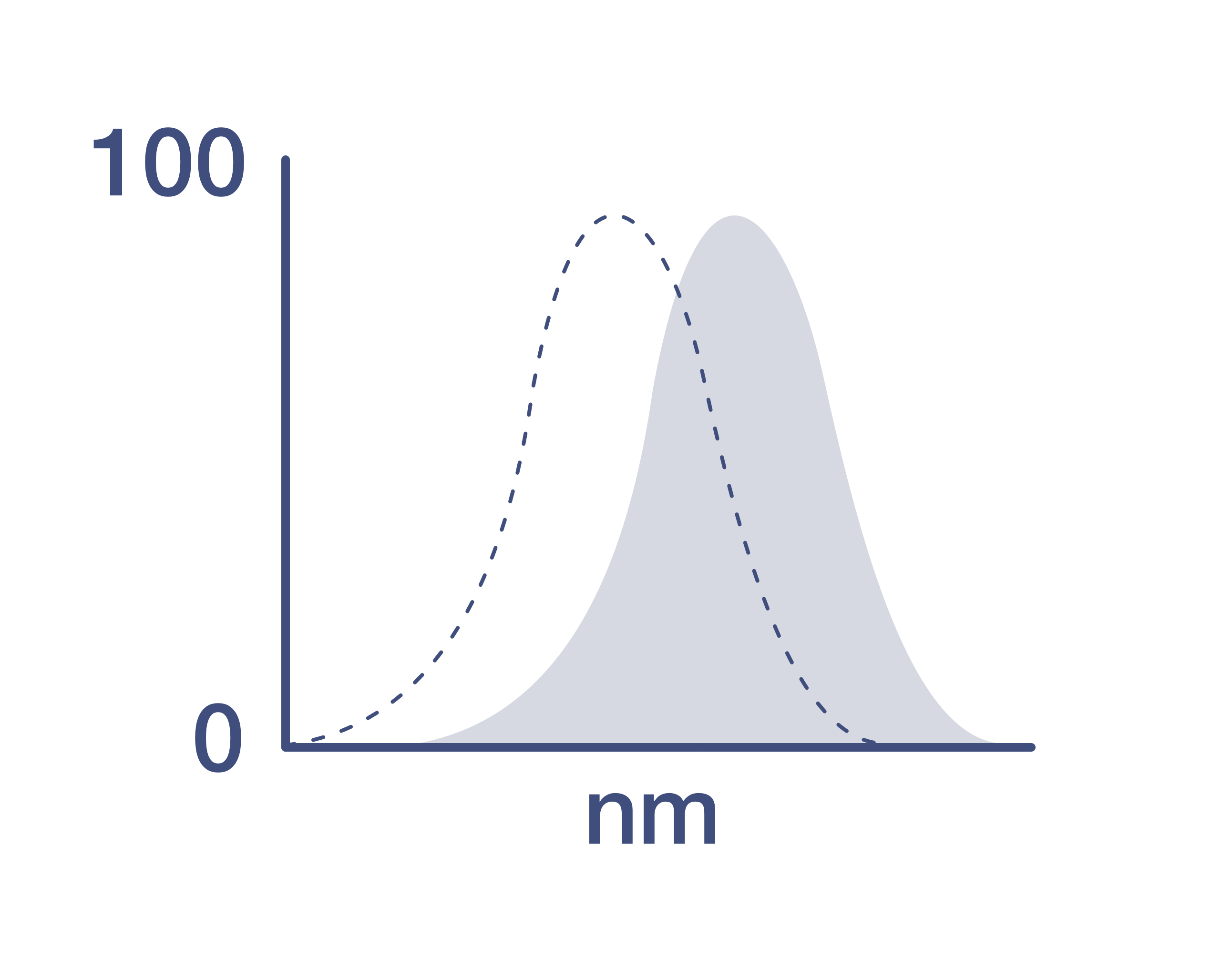
Excitation/Emission Max
492/665 nm
View spectra
Form
Liquid
Concentration
0.2 µg/Test
Purification
Affinity chromatography
Storage buffer
PBS, pH 7.2
Contains
0.09% sodium azide
Storage conditions
4°C, store in dark, DO NOT FREEZE!
RRID
AB_3098684 (AB_2926059)
Product Specific Information
Description
The monoclonal antibody P1H12 recognizes CD146 also known as MUC18, s-endo, Endo-CAM and Mel-CAM, which is a member of the Ig superfamily of proteins. The antibody P1H12 has been reported to crossreact to mouse, rabbit, canine, but not rat.
This product contains 1 vial of NovaFluor conjugate and 1 vial of CellBlox Plus Blocking Buffer.
Applications Tested
This P1H12 antibody has been pre-titrated and tested by flow cytometric analysis of normal human peripheral blood cells and Human Umbilical Vein Cells (HUVEC). This can be used at 5 µL (0.02 µg) per test. A test is defined as the amount (µg) of antibody that will stain a cell sample in a final volume of 100 µL. Cell number should be determined empirically but can range from 10^5 to 10^8 cells/test.
Master mixes
• Master mixes of NFs should be made at 2-8 °C and may be made up to 4 hours ahead of time.
• We do not recommend storing master mixes containing NovaFluor conjugates overnight or longer.
Whole Blood compatibility
• When utilizing whole blood (as opposed to density-gradient-purified PBMC), we recommend lysing red blood cells in bulk prior to staining with NovaFluor conjugates.
• See the Bulk Lysis of Human Whole Blood protocol here.
• Staining of whole blood with NovaFluor conjugates followed by lysis of red blood cells may result in higher-than-expected background staining.
Viability dye compatibility
• NovaFluor dyes are not compatible with DNA intercalating viability dyes.
• Do not use viability dyes such as propidium iodide, 7-actinomycin D (7-AAD) and DAPI. Invitrogen LIVE/DEAD Fixable Dead Cell stains are recommended for use with NovaFluor dyes.
CellBlox Plus Blocking Buffer
• This NovaFluor conjugate comes with CellBlox Plus Blocking Buffer (Cat. No. C001T03F01), essential for optimal staining.
• Use CellBlox Plus Blocking Buffer in all experiments with NovaFluor conjugates.
• Add 5 μL per sample to antibody cocktails/master mixes (regardless of how many Novafluor-conjugated antibodies are present) before combining with cells.
• CellBlox Plus Blocking Buffer is compatible with either Super Bright Complete Blocking Buffer or Brilliant Stain Buffer and can be used in antibody cocktails/master mixes with those reagents.
• For single-color controls, use 5 μL of CellBlox Plus Blocking Buffer per 100 μL of cell sample (10^3 to 10^8 cells).
NovaFluor conjugates are based on Phiton technology utilizing novel fluorophore-containing nucleic acid dye structures that allow for engineered fluorescent signatures with consideration for spillover and spread impacts. Learn more
Excitation: 508 nm; Emission: 664 nm; Laser: 488 nm (Blue) Laser
Target Information
CD146, also known as MCAM (Melanoma Cell Adhesion Molecule), is an integral membrane glycoprotein belonging to the immunoglobulin superfamily. It is heavily glycosylated, with more than 50% of its mass derived from carbohydrates. CD146 is primarily expressed on endothelial cells at cell-cell junctions, playing a crucial role in cell adhesion and the cohesion of the endothelial monolayer in vascular tissue. In addition to endothelial cells, CD146 is expressed on bone marrow fibroblasts, mesenchymal stromal cells, endometrial stromal cells, and some tumors, particularly melanoma. It has also been observed on a subset of circulating T cells and activated lymphocytes, but not on circulating endothelial cells, as indicated by the absence of other endothelial markers such as CD31 and CD51/61. CD146 functions as a calcium-independent cell adhesion molecule and may also act as a signal transduction molecule. It is involved in the recruitment of Fyn kinase and the subsequent tyrosine phosphorylation of intracellular proteins, which are important for actin cytoskeleton assembly. This signaling role includes triggering tyrosine phosphorylation of FYN and PTK2 and causing a transient increase in intracellular calcium concentration. In the context of cancer, CD146 expression may facilitate the interaction of melanoma cells with the vascular system, enhancing hematogenous tumor spread. Additionally, CD146 could serve as an adhesion molecule active in neural crest cells during embryonic development, highlighting its diverse roles in both normal physiological processes and disease states.
For Research Use Only. Not for use in diagnostic procedures. Not for resale without express authorization.
How to use the Panel Builder
Watch the video to learn how to use the Invitrogen Flow Cytometry Panel Builder to build your next flow cytometry panel in 5 easy steps.
References (0)
Have you cited this product in a publication?
Let us know so we can reference it here.
Bioinformatics
Protein Aliases: CD146; Cell surface glycoprotein MUC18; Cell surface glycoprotein P1H12; Gicerin; l-gicerin protein; melanoma adhesion molecule; Melanoma cell adhesion molecule; Melanoma-associated antigen A32; Melanoma-associated antigen MUC18; S-endo 1 endothelial-associated antigen
Gene Aliases: 1-gicerin; AV025631; CD146; CD149; MCAM; MUC18; s-endo; s-gicerin
UniProt ID: (Human) P43121, (Mouse) Q8R2Y2
Entrez Gene ID: (Human) 4162, (Dog) 489368, (Mouse) 84004

Performance Guarantee
If an Invitrogen™ antibody doesn't perform as described on our website or datasheet,we'll replace the product at no cost to you, or provide you with a credit for a future purchase.*
Learn more
We're here to help
Get expert recommendations for common problems or connect directly with an on staff expert for technical assistance related to applications, equipment and general product use.
Contact tech support