Search Thermo Fisher Scientific
Disclaimer

Clicking the images or links will redirect you to a website hosted by BenchSci that provides third-party scientific content. Neither the content nor the BenchSci technology and processes for selection have been evaluated by us; we are providing them as-is and without warranty of any kind, including for use or application of the Thermo Fisher Scientific products presented.
Invitrogen
CD1c Monoclonal Antibody (L161), NovaFluor™ Blue 610-70S, eBioscience™
{{$productOrderCtrl.translations['antibody.pdp.commerceCard.promotion.promotions']}}
{{$productOrderCtrl.translations['antibody.pdp.commerceCard.promotion.viewpromo']}}
{{$productOrderCtrl.translations['antibody.pdp.commerceCard.promotion.promocode']}}: {{promo.promoCode}} {{promo.promoTitle}} {{promo.promoDescription}}. {{$productOrderCtrl.translations['antibody.pdp.commerceCard.promotion.learnmore']}}
")
FIGURE: 1 / 2
CD1c Antibody (H046T03B06-A) in Flow
Normal human peripheral blood cells were unstained (left) or stained with CD1c Monoclonal Antibody, NovaFluor Blue 610-70S (right). All cells were co-stained with CD19 Monoclonal Antibody, eFluor 450 (Product # 48-0199-42). Total viable cells in the lymphocyte gate were used for analysis, as determined by LIVE/DEAD Blue (Product # L34962). Data was acquired on a 5-laser Cytek Aurora and unmixed with autofluorescence extraction.


 in Flow")
 in Flow")
Product Details
H046T03B06-A
Product Specifications
Species Reactivity
Human
Host/Isotype
Mouse
/ IgG1, kappa
Class
Monoclonal
Type
Antibody
Clone
L161
Conjugate
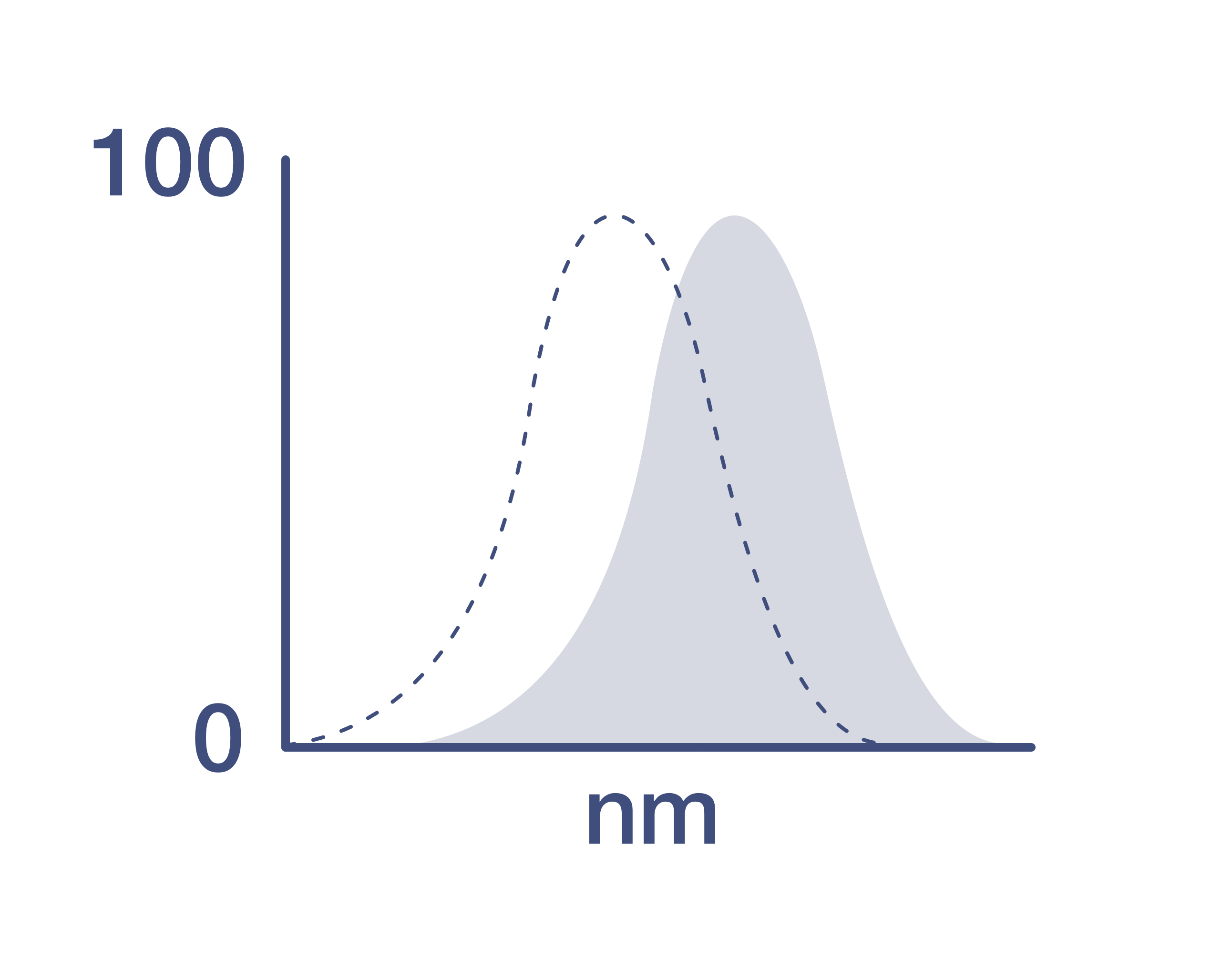
Excitation/Emission Max
492/616 nm
View spectra
Form
Liquid
Concentration
0.2 µg/Test
Purification
Affinity chromatography
Storage buffer
PBS, pH 7.2
Contains
0.09% sodium azide
Storage conditions
4°C, store in dark, DO NOT FREEZE!
RRID
AB_3098560 (AB_2925949)
Product Specific Information
Description
This L161 monoclonal antibody detects CD1c (also known as BDCA-1), a glycoprotein that is noncovalently linked to beta-2 microglobulin on thymocytes and antigen presenting cells such as dendritic and Langerhans cells.
This product contains 1 vial of NovaFluor conjugate and 1 vial of CellBlox Plus Blocking Buffer.
Applications Tested
This L161 antibody has been pre-titrated and tested by flow cytometric analysis of normal human peripheral blood cells. This can be used at 5 µL (0.06 µg) per test. A test is defined as the amount (µg) of antibody that will stain a cell sample in a final volume of 100 µL. Cell number should be determined empirically but can range from 10^5 to 10^8 cells/test.
Master mixes
• Master mixes of NFs should be made at 2-8 °C and may be made up to 4 hours ahead of time.
• We do not recommend storing master mixes containing NovaFluor conjugates overnight or longer.
Whole Blood compatibility
• When utilizing whole blood (as opposed to density-gradient-purified PBMC), we recommend lysing red blood cells in bulk prior to staining with NovaFluor conjugates.
• See the Bulk Lysis of Human Whole Blood protocol here.
• Staining of whole blood with NovaFluor conjugates followed by lysis of red blood cells may result in higher-than-expected background staining.
Viability dye compatibility
• NovaFluor dyes are not compatible with DNA intercalating viability dyes.
• Do not use viability dyes such as propidium iodide, 7-actinomycin D (7-AAD) and DAPI. Invitrogen LIVE/DEAD Fixable Dead Cell stains are recommended for use with NovaFluor dyes.
CellBlox Plus Blocking Buffer
• This NovaFluor conjugate comes with CellBlox Plus Blocking Buffer (Cat. No. C001T03F01), essential for optimal staining.
• Use CellBlox Plus Blocking Buffer in all experiments with NovaFluor conjugates.
• Add 5 μL per sample to antibody cocktails/master mixes (regardless of how many Novafluor-conjugated antibodies are present) before combining with cells.
• CellBlox Plus Blocking Buffer is compatible with either Super Bright Complete Blocking Buffer or Brilliant Stain Buffer and can be used in antibody cocktails/master mixes with those reagents.
• For single-color controls, use 5 μL of CellBlox Plus Blocking Buffer per 100 μL of cell sample (10^3 to 10^8 cells).
NovaFluor conjugates are based on Phiton technology utilizing novel fluorophore-containing nucleic acid dye structures that allow for engineered fluorescent signatures with consideration for spillover and spread impacts. Learn more
Excitation: 492 nm; Emission: 616 nm; Laser: 488 nm (Blue) Laser
Target Information
CD1c is a member of the CD1 family of transmembrane glycoproteins, which are structurally related to major histocompatibility complex (MHC) proteins and form heterodimers with beta-2-microglobulin. This family of proteins is involved in the presentation of lipid and glycolipid antigens, both of self and microbial origin, to T cells during the adaptive immune response. CD1c is expressed on some circulating and marginal zone B cells, as well as in lymph nodes and germinal centers. It plays a crucial role in presenting lipid antigens, such as microbial fatty acids, to effector T cells. The protein encoded by the CD1c gene localizes to late endosomes and lysosomes, utilizing a tyrosine-based motif in its cytoplasmic tail for targeting. Vesicular acidification is required for CD1c to bind lipid antigens effectively. The human genome contains five CD1 family genes organized in a cluster on chromosome 1, with each member differing in cellular localization and specificity for particular lipid ligands. CD1c undergoes alternative splicing, resulting in three different isoforms: soluble, membrane-bound, and cytoplasmic/soluble isoforms, highlighting its functional diversity in immune processes.
For Research Use Only. Not for use in diagnostic procedures. Not for resale without express authorization.
How to use the Panel Builder
Watch the video to learn how to use the Invitrogen Flow Cytometry Panel Builder to build your next flow cytometry panel in 5 easy steps.
References (0)
Have you cited this product in a publication?
Let us know so we can reference it here.
Bioinformatics
Protein Aliases: CD1c; CD1C antigen, c polypeptide; cortical thymocyte antigen CD1C; differentiation antigen CD1-alpha-3; RP11-101J8.3; T-cell surface glycoprotein CD1c
Gene Aliases: BDCA1; CD1; CD1A; CD1C; R7
UniProt ID: (Human) P29017
Entrez Gene ID: (Human) 911

Performance Guarantee
If an Invitrogen™ antibody doesn't perform as described on our website or datasheet,we'll replace the product at no cost to you, or provide you with a credit for a future purchase.*
Learn more
We're here to help
Get expert recommendations for common problems or connect directly with an on staff expert for technical assistance related to applications, equipment and general product use.
Contact tech support