Search Thermo Fisher Scientific
Disclaimer

Clicking the images or links will redirect you to a website hosted by BenchSci that provides third-party scientific content. Neither the content nor the BenchSci technology and processes for selection have been evaluated by us; we are providing them as-is and without warranty of any kind, including for use or application of the Thermo Fisher Scientific products presented.
Invitrogen
CD235a (Glycophorin A) Monoclonal Antibody (10F7MN), Brilliant Ultra Violet™ 737, eBioscience™
{{$productOrderCtrl.translations['antibody.pdp.commerceCard.promotion.promotions']}}
{{$productOrderCtrl.translations['antibody.pdp.commerceCard.promotion.viewpromo']}}
{{$productOrderCtrl.translations['antibody.pdp.commerceCard.promotion.promocode']}}: {{promo.promoCode}} {{promo.promoTitle}} {{promo.promoDescription}}. {{$productOrderCtrl.translations['antibody.pdp.commerceCard.promotion.learnmore']}}
 Antibody in Flow Cytometry (Flow)")
FIGURE: 1 / 1
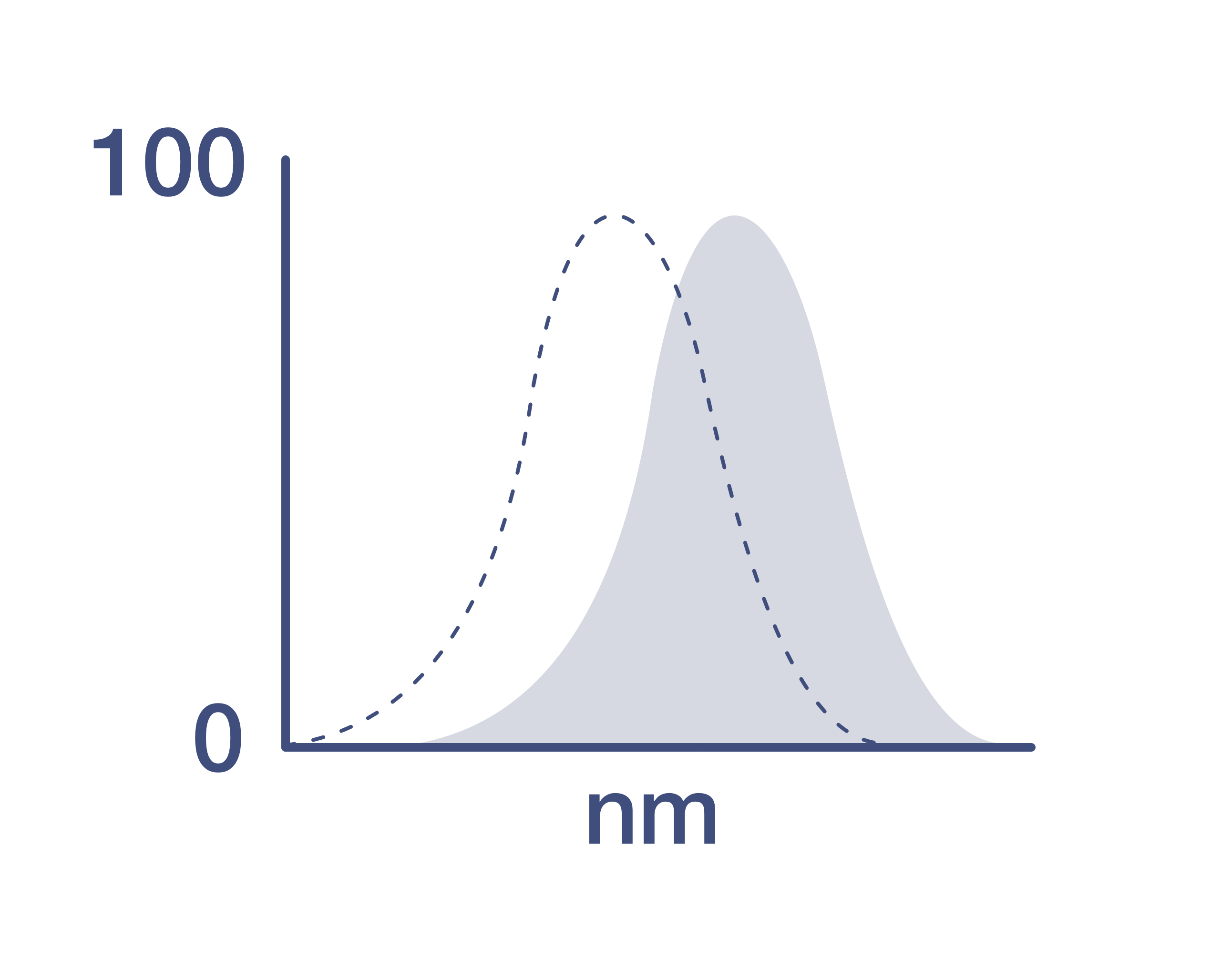
CD235a (Glycophorin A) Antibody (367-9886-42) in Flow
Normal human peripheral blood cells were stained with Mouse IgG1 kappa Isotype Control, Brilliant Ultra Violet 737 (BUV737) (Product # 367-4714-81) (blue histogram) or CD235a (Glycophorin A) Monoclonal Antibody, Brilliant Ultra Violet 737 (BUV737) (purple histogram). Cells in the erythrocyte gate were used for analysis.

 Antibody (367-9886-42) in Flow")
Product Details
367-9886-42
Product Specifications
Species Reactivity
Human
Host/Isotype
Mouse
/ IgG1, kappa
Recommended Isotype Control
Class
Monoclonal
Type
Antibody
Clone
10F7MN
Conjugate
Excitation/Emission Max
350/740 nm
View spectra
Form
Liquid
Concentration
5 µL/Test
Purification
Affinity chromatography
Storage buffer
PBS, pH 7.2, with BSA
Contains
0.09% sodium azide
Storage conditions
4°C, store in dark, DO NOT FREEZE!
Shipping conditions
Ambient (domestic); Wet ice (international)
RRID
AB_2896056
Product Specific Information
Description: The monoclonal antibody 10F7MN recognizes human glycophorin A (also known as CD235a). The antibody can see both the M and N alleles. Glycophorin A is a 151 amino acid sialoglycoprotein found on the erythrocyte (RBC) and erthyroid progenitor cell membrane at about 500,000 copies per cell. The gene for glycophorin resides on chromosome 4 and has 2 allelic forms: M and N, which differ in two amino acids. The M group possesses Ser1 and Gly5 while the N group has Leu1 and Glu5. Recent data suggest that exposure to toxins can cause mutation or loss of an allele resulting in phenotypic changes. Studies are also beginning to correlate genotype/phenotype with predisposition to cancer and heart disease.
Applications Reported: This 10F7MN antibody has been reported for use in flow cytometric analysis.
Applications Tested: This 10F7MN antibody has been pre-diluted and tested by flow cytometric analysis of normal human peripheral blood cells. This may be used at 5 µL (0.25 µg) per test. A test is defined as the amount (µg) of antibody that will stain a cell sample in a final volume of 100 µL. Cell number should be determined empirically but can range from 10^5 to 10^8 cells/test.
Brilliant Ultra Violet™ 737 (BUV737) is a tandem dye that emits at 732 nm and is intended for use on cytometers equipped with an ultraviolet (355 nm) laser. Please make sure that your instrument is capable of detecting this fluorochrome.
When using two or more Super Bright, Brilliant Violet™, Brilliant Ultra Violet™, or other polymer dye-conjugated antibodies in a staining panel, it is recommended to use Super Bright Complete Staining Buffer (Product # SB-4401) or Brilliant Stain Buffer (Product # 00-4409) to minimize any non-specific polymer interactions. Please refer to the datasheet for Super Bright Staining Buffer or Brilliant Stain Buffer for more information.
Light sensitivity: This tandem dye is sensitive to photo-induced oxidation. Please protect this vial and stained samples from light.
Fixation: Samples can be stored in IC Fixation Buffer (Product # 00-8222) (100 µL of cell sample + 100 µL of IC Fixation Buffer) or 1-step Fix/Lyse Solution (Product # 00-5333) for up to 3 days in the dark at 4°C with minimal impact on brightness and FRET efficiency/compensation. Some generalizations regarding fluorophore performance after fixation can be made, but clone specific performance should be determined empirically.
Our internal testing suggests that Brilliant Ultra Violet™ 737 (BUV737) is compatible with short-term methanol-based fixation, but should not be stored in buffers containing methanol for longer than one hour.
Excitation: 355 nm; Emission: 732 nm; Laser: Ultraviolet Laser.
BRILLIANT ULTRA VIOLET™ is a trademark or registered trademark of Becton, Dickinson and Company or its affiliates, and is used under license. Powered by Sirigen™.
Target Information
Glycophorin A, also known as CD235a, is a 151 amino acid sialoglycoprotein expressed on the membrane of mature erythrocytes and erythroid progenitor cells, with approximately 500,000 copies per cell. The gene for glycophorin A is located on chromosome 4 and exists in two allelic forms, M and N, which differ by two amino acids: the M group has Ser1 and Gly5, while the N group has Leu1 and Glu5. These allelic variations define the blood group M and N specificities. Glycophorin A serves multiple functions, including providing a mucin-like barrier that minimizes aggregation between red blood cells in circulation, potentially preventing cell fusion. It also acts as a receptor for certain pathogens, including Sandei virus, parvovirus, and Hsa, a Streptococcus adhesin. Recent studies suggest that exposure to toxins can lead to mutations or loss of alleles, resulting in phenotypic changes. There is ongoing research correlating genotype and phenotype with predisposition to diseases such as cancer and heart disease, highlighting the importance of glycophorin A in both health and disease. Glycophorin A is a significant marker for studying erythrocyte-related functions and pathologies, as well as its role in pathogen interactions and disease predisposition.
For Research Use Only. Not for use in diagnostic procedures. Not for resale without express authorization.
How to use the Panel Builder
Watch the video to learn how to use the Invitrogen Flow Cytometry Panel Builder to build your next flow cytometry panel in 5 easy steps.
References (0)
Have you cited this product in a publication?
Let us know so we can reference it here.
Bioinformatics
Protein Aliases: CD235; CD235a; erythroid-lineage-specific membrane sialoglycoprotein; glycophorin A (MN blood group); glycophorin A, GPA; glycophorin Erik; glycophorin MiI; glycophorin MiV; glycophorin SAT; glycophorin Sta type C; Glycophorin-A; HGNC:4702; HGpMiX; HGpStaC; Mi.V glycoprotein; Mi.V glycoprotein (24 AA); MN sialoglycoprotein; PAS-2; recombinant glycophorin A-B Miltenberger-DR; Sialoglycoprotein alpha
Gene Aliases: CD235a; GPA; GPErik; GPSAT; GYPA; HGpMiV; HGpMiXI; HGpSta(C); MN; MNS; PAS-2
UniProt ID: (Human) P02724
Entrez Gene ID: (Human) 2993

Performance Guarantee
If an Invitrogen™ antibody doesn't perform as described on our website or datasheet,we'll replace the product at no cost to you, or provide you with a credit for a future purchase.*
Learn more
We're here to help
Get expert recommendations for common problems or connect directly with an on staff expert for technical assistance related to applications, equipment and general product use.
Contact tech support