Search Thermo Fisher Scientific
Disclaimer

Clicking the images or links will redirect you to a website hosted by BenchSci that provides third-party scientific content. Neither the content nor the BenchSci technology and processes for selection have been evaluated by us; we are providing them as-is and without warranty of any kind, including for use or application of the Thermo Fisher Scientific products presented.
Invitrogen
CD41a Monoclonal Antibody (eBioMWReg30 (MWReg30)), NovaFluor™ Yellow 660, eBioscience™
{{$productOrderCtrl.translations['antibody.pdp.commerceCard.promotion.promotions']}}
{{$productOrderCtrl.translations['antibody.pdp.commerceCard.promotion.viewpromo']}}
{{$productOrderCtrl.translations['antibody.pdp.commerceCard.promotion.promocode']}}: {{promo.promoCode}} {{promo.promoTitle}} {{promo.promoDescription}}. {{$productOrderCtrl.translations['antibody.pdp.commerceCard.promotion.learnmore']}}
")
FIGURE: 1 / 3
CD41a Antibody (M044T03Y04-A) in Flow
Mouse platelets were either left unstained (blue histogram) or stained with 0.6 µg of CD41a Monoclonal Antibody, NovaFluor Yellow 660 (purple histogram). Total viable platelets in the platelet gate were used for analysis, as determined by LIVE/DEAD Blue (Product # L34962). Data was acquired on a 5-laser Cytek Aurora and unmixed with autofluorescence extraction.


 in Flow")
 in Flow")
 in Flow")
Product Details
M044T03Y04-A
Product Specifications
Species Reactivity
Mouse
Host/Isotype
Rat
/ IgG1, kappa
Class
Monoclonal
Type
Antibody
Clone
eBioMWReg30 (MWReg30)
Conjugate
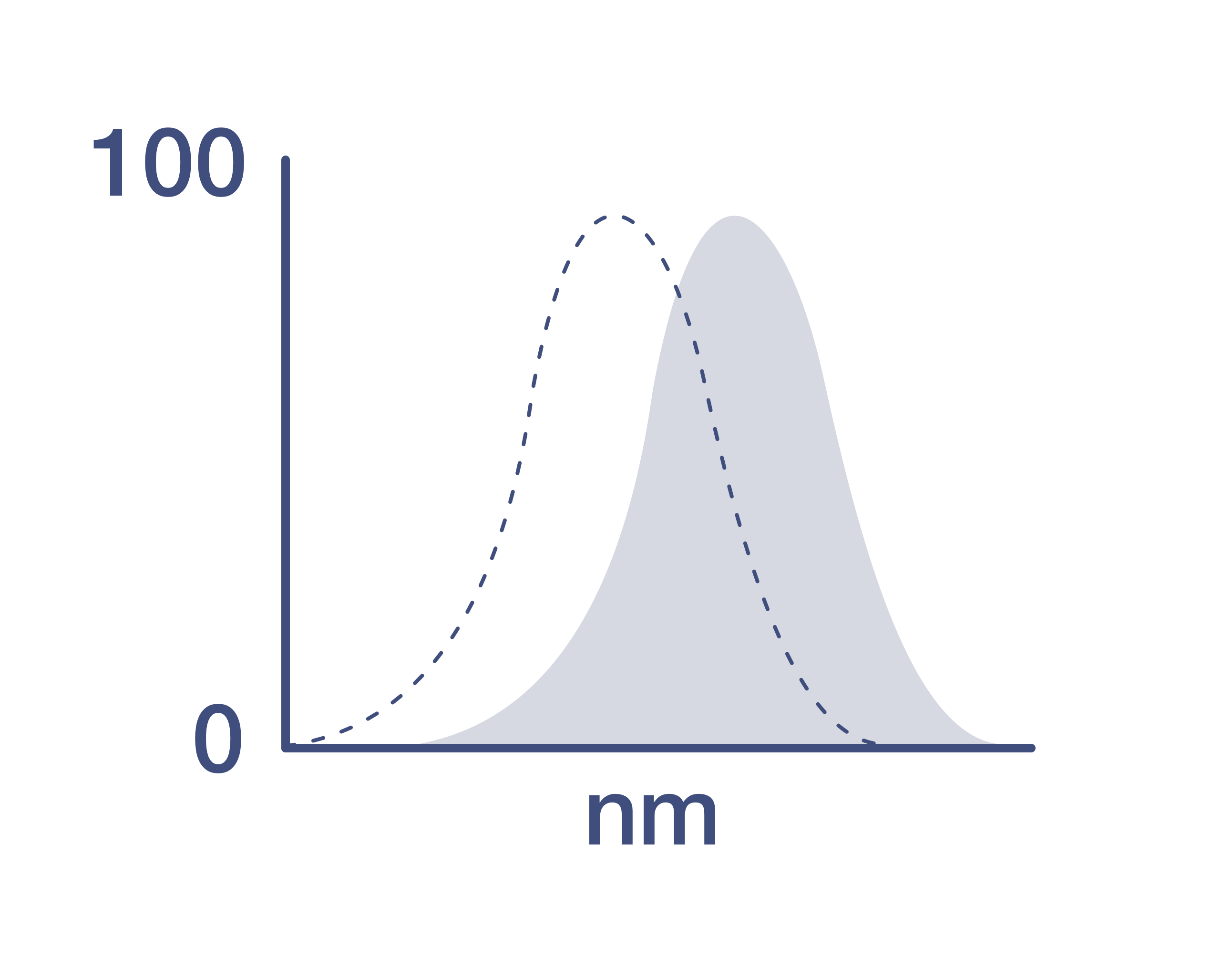
Excitation/Emission Max
550/664 nm
View spectra
Form
Liquid
Concentration
0.1 mg/mL
Purification
Affinity chromatography
Storage buffer
PBS, pH 7.2
Contains
0.09% sodium azide
Storage conditions
4°C, store in dark, DO NOT FREEZE!
RRID
AB_3099169 (AB_2926288)
Product Specific Information
Description
The eBioMWReg30 monoclonal antibody reacts with mouse CD41 (fibrinogen receptor, gpIIb, integrin alpha Iib). Recently, the SLAM-family markers, CD48 and CD150 have been used to reliably identify hematopoietic stem cells (HSC). Specifically, it was found that CD150+CD48- bone marrow cells were highly efficient in their ability to confer long-term multi-lineage reconstitution in irradiated mice. Furthermore, the efficiency of reconstitution was enhanced when HSCs were further enriched through the exclusion of CD41+ cells. Thus, the use of CD150+CD48-CD41- as an expression profile efficiently identifies hematopoietic stem cells.
This product contains 1 vial of NovaFluor conjugate and 1 vial of CellBlox Plus Blocking Buffer.
Applications Tested
This eBioMWReg30 (MWReg30) antibody has been tested by flow cytometric analysis of mouse platelets. This can be used at less than or equal to 0.125 µg per test. A test is defined as the amount (µg) of antibody that will stain a cell sample in a final volume of 100 µL. Cell number should be determined empirically but can range from 10^5 to 10^8 cells/test. It is recommended that the antibody be carefully titrated for optimal performance in the assay of interest.
Master mixes
• Master mixes of NFs should be made at 2-8 °C and may be made up to 4 hours ahead of time.
• We do not recommend storing master mixes containing NovaFluor conjugates overnight or longer.
Whole Blood compatibility
• When utilizing whole blood (as opposed to density-gradient-purified PBMC), we recommend lysing red blood cells in bulk prior to staining with NovaFluor conjugates.
• See the Bulk Lysis of Human Whole Blood protocol here.
• Staining of whole blood with NovaFluor conjugates followed by lysis of red blood cells may result in higher-than-expected background staining.
Viability dye compatibility
• NovaFluor dyes are not compatible with DNA intercalating viability dyes.
• Do not use viability dyes such as propidium iodide, 7-actinomycin D (7-AAD) and DAPI. Invitrogen LIVE/DEAD Fixable Dead Cell stains are recommended for use with NovaFluor dyes.
CellBlox Plus Blocking Buffer
• This NovaFluor conjugate comes with CellBlox Plus Blocking Buffer (Cat. No. C001T03F01), essential for optimal staining.
• Use CellBlox Plus Blocking Buffer in all experiments with NovaFluor conjugates.
• Add 5 μL per sample to antibody cocktails/master mixes (regardless of how many Novafluor-conjugated antibodies are present) before combining with cells.
• CellBlox Plus Blocking Buffer is compatible with either Super Bright Complete Blocking Buffer or Brilliant Stain Buffer and can be used in antibody cocktails/master mixes with those reagents.
• For single-color controls, use 5 μL of CellBlox Plus Blocking Buffer per 100 μL of cell sample (10^3 to 10^8 cells).
NovaFluor conjugates are based on Phiton technology utilizing novel fluorophore-containing nucleic acid dye structures that allow for engineered fluorescent signatures with consideration for spillover and spread impacts. Learn more
Our internal testing shows that NovaFluor Yellow 660 non-specifically stains B cells in SJL mice. Non-specific staining has not been observed in BALB/c or C57BL/6 mice. Other strains have not been tested. See the Antibody Testing Data for an example of this strain-dependent difference.
Excitation: 550 nm; Emission: 664 nm; Laser: 561 nm (Yellow) Laser
Target Information
CD41, also known as platelet glycoprotein IIb or ITGA2B, is a protein composed of two subunits: a 120 kDa alpha subunit and a 23 kDa beta subunit. It interacts with CD61 (gpIIIa, integrin beta III) in the presence of calcium to form a functional adhesive protein receptor. Initially thought to be expressed exclusively on platelets and megakaryocytes, CD41 is also found on hematopoietic progenitors in the embryo, fetus, and adult, indicating its role in early stages of hematopoietic differentiation. CD41 plays a crucial role in platelet function and blood coagulation by mediating platelet aggregation. Upon blood vessel damage, CD41 binds to various adhesion molecules, including von Willebrand factor, fibrinogen, fibronectin, and vitronectin, facilitating platelet adhesion and aggregation. This receptor is essential for platelet function, contributing to the binding of several adhesion molecules and playing a significant role in hemostasis. Diseases associated with CD41 dysfunction include Glanzmann Thrombasthenia and Platelet type-16 bleeding disorder, highlighting its importance in normal platelet function and blood coagulation processes.
For Research Use Only. Not for use in diagnostic procedures. Not for resale without express authorization.
How to use the Panel Builder
Watch the video to learn how to use the Invitrogen Flow Cytometry Panel Builder to build your next flow cytometry panel in 5 easy steps.
References (0)
Have you cited this product in a publication?
Let us know so we can reference it here.
Bioinformatics
Protein Aliases: alpha IIb; CD41; form 1; form 2; GP IIb; GPalpha IIb; GPIIb; Integrin alpha-IIb; platelet glycoprotein IIb of IIb/II Ia complex; Platelet membrane glycoprotein IIb
Gene Aliases: AI172977; alphaIIb; CD41; CD41B; GpIIb; Itga2b
UniProt ID: (Mouse) Q9QUM0
Entrez Gene ID: (Mouse) 16399

Performance Guarantee
If an Invitrogen™ antibody doesn't perform as described on our website or datasheet,we'll replace the product at no cost to you, or provide you with a credit for a future purchase.*
Learn more
We're here to help
Get expert recommendations for common problems or connect directly with an on staff expert for technical assistance related to applications, equipment and general product use.
Contact tech support