Search Thermo Fisher Scientific
Disclaimer

Clicking the images or links will redirect you to a website hosted by BenchSci that provides third-party scientific content. Neither the content nor the BenchSci technology and processes for selection have been evaluated by us; we are providing them as-is and without warranty of any kind, including for use or application of the Thermo Fisher Scientific products presented.
Invitrogen
CD8a Monoclonal Antibody (53-6.7), Brilliant Ultra Violet™ 615, eBioscience™
{{$productOrderCtrl.translations['antibody.pdp.commerceCard.promotion.promotions']}}
{{$productOrderCtrl.translations['antibody.pdp.commerceCard.promotion.viewpromo']}}
{{$productOrderCtrl.translations['antibody.pdp.commerceCard.promotion.promocode']}}: {{promo.promoCode}} {{promo.promoDescription}}. {{$productOrderCtrl.translations['antibody.pdp.commerceCard.promotion.learnmore']}}
")
FIGURE: 1 / 1
CD8a Antibody (366-0081-82) in Flow
C57BL/6 mouse splenocytes were stained with CD4 Monoclonal Antibody, FITC (Product # 11-0042-82) and 1.0 µg of Rat IgG2a kappa Isotype Control, Brilliant Ultra Violet 615 (Product # 366-4321-81) (left) or 1.0 µg of CD8a Monoclonal Antibody, Brilliant Ultra Violet 615 (right). Viable cells in the lymphocyte gate were used for analysis, as determined by 7-AAD (Product # 00-6993-50).

 in Flow")
Product Details
366-0081-82
Product Specifications
Species Reactivity
Mouse
Host/Isotype
Rat
/ IgG2a, kappa
Recommended Isotype Control
Class
Monoclonal
Type
Antibody
Clone
53-6.7
Conjugate
Excitation/Emission Max
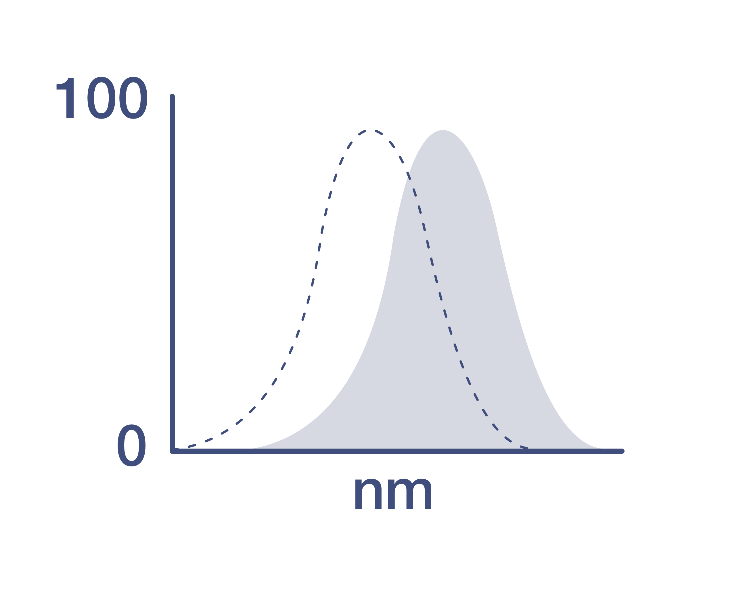
350/607 nm
View spectra
Form
Liquid
Concentration
0.2 mg/mL
Purification
Affinity chromatography
Storage buffer
PBS, pH 7.2, with BSA
Contains
0.09% sodium azide
Storage conditions
4°C, store in dark, DO NOT FREEZE!
Shipping conditions
Ambient (domestic); Wet ice (international)
RRID
AB_2920984
Product Specific Information
Description: The 53-6.7 monoclonal antibody reacts with the mouse CD8a molecule. CD8a is an approximately 32-34 kDa cell surface receptor expressed either as a heterodimer with the CD8 beta chain (CD8 alpha beta) or as a homodimer (CD8 alpha alpha). A majority of thymocytes and a subpopulation of mature alpha beta TCR T cells express CD8 alpha beta while gamma delta TCR T cells, a subpopulation of intestinal intraepithelial lymphocytes (IELs) and dendritic cells express CD8 alpha alpha. CD8 binds to MHC class I and through its association with protein tyrosine kinase p56lck plays a role in T cell development and activation of mature T cells.
Applications Reported: This 53-6.7 antibody has been reported for use in flow cytometric analysis.
Applications Tested: This 53-6.7 antibody has been tested by flow cytometric analysis of mouse splenocytes. This may be used at less than or equal to 1.0 µg per test. A test is defined as the amount (µg) of antibody that will stain a cell sample in a final volume of 100 µL. Cell number should be determined empirically but can range from 10^5 to 10^8 cells/test. It is recommended that the antibody be carefully titrated for optimal performance in the assay of interest.
Brilliant Ultra Violet™ 615 (BUV615) is a tandem dye that emits at 615 nm and is intended for use on cytometers equipped with an ultraviolet (355 nm) laser. Please make sure that your instrument is capable of detecting this fluorochrome.
When using two or more Super Bright, Brilliant Violet™, Brilliant Ultra Violet™, or other polymer dye-conjugated antibodies in a staining panel, it is recommended to use Super Bright Complete Staining Buffer (Product # SB-4401-42) or Brilliant Stain Buffer™ (Product # 00-4409-75) to minimize any non-specific polymer interactions. Please refer to the datasheet for Super Bright Staining Buffer or Brilliant Stain Buffer for more information.
Light sensitivity: This tandem dye is sensitive to photo-induced oxidation. Please protect this vial and stained samples from light.
Fixation: Samples can be stored in IC Fixation Buffer (Product # 00-8222-49) (100 µL of cell sample + 100 µL of IC Fixation Buffer) or 1-step Fix/Lyse Solution (Product # 00-5333-54) for up to 3 days in the dark at 4°C with minimal impact on brightness and FRET efficiency/compensation. Some generalizations regarding fluorophore performance after fixation can be made, but clone-specific performance should be determined empirically.
Excitation: 350 nm; Emission: 615 nm; Laser: Ultraviolet Laser.
BRILLIANT ULTRA VIOLET™ is a trademark or registered trademark of Becton, Dickinson and Company or its affiliates, and is used under license. Powered by Sirigen™.
Target Information
CD8, also known as cluster of differentiation 8, is a type I transmembrane glycoprotein of the immunoglobulin family that plays a crucial role in T cell differentiation, activation, and signal transduction. It is expressed as either a heterodimer (CD8 alpha beta) or a homodimer (CD8 alpha alpha). The CD8 alpha beta form is predominantly found on the majority of thymocytes and a subpopulation of mature alpha beta TCR T cells, while the CD8 alpha alpha form is expressed on gamma delta TCR T cells, a subset of intestinal intraepithelial lymphocytes (IELs), and dendritic cells. CD8 functions as a co-receptor for major histocompatibility complex class I (MHC-I) molecules, working alongside the T cell receptor (TCR). The CD8 alpha chain is essential for binding to MHC-I. CD8 is also expressed on a subset of T cells, NK cells, monocytes, and dendritic cells as disulfide-linked homodimers of CD8 alpha. Upon ligation of MHC-I/peptide complexes presented by antigen-presenting cells (APCs), CD8 recruits lymphocyte-specific protein tyrosine kinase (Lck), leading to lymphokine production, increased motility, and activation of cytotoxic T lymphocytes (CTLs). Activated CTLs are vital for clearing pathogens and tumor cells. The differentiation of naive CD8+ T cells into CTLs is strongly enhanced by cytokines such as IL-2, IL-12, and TGF-beta1. Through its interactions with MHC-I and association with protein tyrosine kinase p56lck, CD8 plays a significant role in T cell development and the activation of mature T cells.
For Research Use Only. Not for use in diagnostic procedures. Not for resale without express authorization.
How to use the Panel Builder
Watch the video to learn how to use the Invitrogen Flow Cytometry Panel Builder to build your next flow cytometry panel in 5 easy steps.
References (0)
Have you cited this product in a publication?
Let us know so we can reference it here.
Bioinformatics
Protein Aliases: CD8a; CD8alpha; CD8b; CD8beta; fCD8; Leu-2; leu-2a; Lymphocyte antigen 3; Lyt-2.1 lymphocyte differentiation antigen (AA at 100); T-cell membrane glycoprotein Ly-3; T-cell surface glycoprotein CD8 alpha chain; T-cell surface glycoprotein CD8 beta chain; T-cell surface glycoprotein Lyt-2; T-cell surface glycoprotein Lyt-3
Gene Aliases: BB154331; Cd8a; Cd8b; Cd8b1; Ly-2; Ly-3; Ly-35; Ly-B; Ly-C; Lyt-2; Lyt-3; Lyt2; Lyt3
UniProt ID: (Mouse) P01731, (Mouse) P10300
Entrez Gene ID: (Mouse) 12525, (Mouse) 12526

Performance Guarantee
If an Invitrogen™ antibody doesn't perform as described on our website or datasheet,we'll replace the product at no cost to you, or provide you with a credit for a future purchase.*
Learn more
We're here to help
Get expert recommendations for common problems or connect directly with an on staff expert for technical assistance related to applications, equipment and general product use.
Contact tech support