Search Thermo Fisher Scientific
Disclaimer

Clicking the images or links will redirect you to a website hosted by BenchSci that provides third-party scientific content. Neither the content nor the BenchSci technology and processes for selection have been evaluated by us; we are providing them as-is and without warranty of any kind, including for use or application of the Thermo Fisher Scientific products presented.
Invitrogen
MHC Class II I-Ab Monoclonal Antibody (AF6-120.1), NovaFluor™ Red 755, eBioscience™
{{$productOrderCtrl.translations['antibody.pdp.commerceCard.promotion.promotions']}}
{{$productOrderCtrl.translations['antibody.pdp.commerceCard.promotion.viewpromo']}}
{{$productOrderCtrl.translations['antibody.pdp.commerceCard.promotion.promocode']}}: {{promo.promoCode}} {{promo.promoTitle}} {{promo.promoDescription}}. {{$productOrderCtrl.translations['antibody.pdp.commerceCard.promotion.learnmore']}}
")
FIGURE: 1 / 2
MHC Class II I-Ab Antibody (M046T03R06-A) in Flow
C57BL/6 mouse splenocytes were unstained (left) or stained with 0.6 µg of MHC Class II I-Ab Monoclonal Antibody, NovaFluor Red 755 (right). All cells were co-stained with CD45R (B220) Monoclonal Antibody, eFluor 450 (Product # 48-0452-82). Total viable cells in the lymphocyte gate were used for analysis, as determined by LIVE/DEAD Blue (Product # L34962). Data was acquired on a 5-laser Cytek Aurora and unmi... View More


 in Flow")
 in Flow")
Product Details
M046T03R06-A
Product Specifications
Species Reactivity
Mouse
Host/Isotype
Mouse
/ IgG2a, kappa
Class
Monoclonal
Type
Antibody
Clone
AF6-120.1
Conjugate
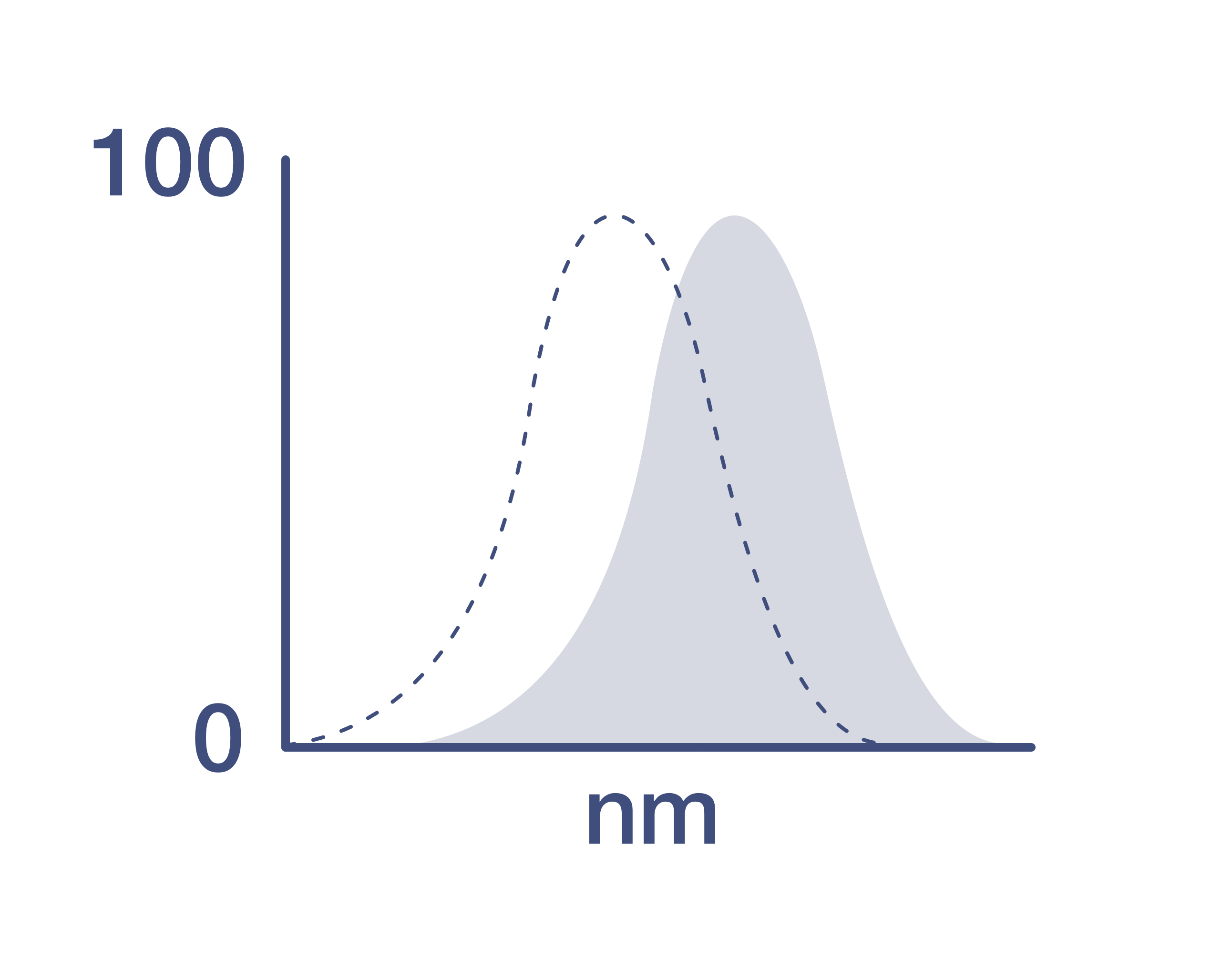
Excitation/Emission Max
636/755 nm
View spectra
Form
Liquid
Concentration
0.1 µg/Test
Purification
Affinity chromatography
Storage buffer
PBS, pH 7.2
Contains
0.09% sodium azide
Storage conditions
4°C, store in dark, DO NOT FREEZE!
RRID
AB_3099185 (AB_2931028)
Product Specific Information
Description
This AF6-120.1.2 monoclonal antibody reacts with the mouse MHC Class II I-Ab alloantigen of H-2b bearing mouse strains, including C57Bl/6 and 129. This cell surface molecule is involved in antigen presentation to T cells expressing CD3/TCR and CD4.
This product contains 1 vial of NovaFluor conjugate and 1 vial of CellBlox Plus Blocking Buffer.
Applications Tested
This AF6-120.1 antibody has been tested by flow cytometric analysis of mouse splenocytes. This can be used at less than or equal to 0.6 µg per test. A test is defined as the amount (µg) of antibody that will stain a cell sample in a final volume of 100 µL. Cell number should be determined empirically but can range from 10^5 to 10^8 cells/test. It is recommended that the antibody be carefully titrated for optimal performance in the assay of interest.
Master mixes
• Master mixes of NFs should be made at 2-8 °C and may be made up to 4 hours ahead of time.
• We do not recommend storing master mixes containing NovaFluor conjugates overnight or longer.
Whole Blood compatibility
• When utilizing whole blood (as opposed to density-gradient-purified PBMC), we recommend lysing red blood cells in bulk prior to staining with NovaFluor conjugates.
• See the Bulk Lysis of Human Whole Blood protocol here.
• Staining of whole blood with NovaFluor conjugates followed by lysis of red blood cells may result in higher-than-expected background staining.
Viability dye compatibility
• NovaFluor dyes are not compatible with DNA intercalating viability dyes.
• Do not use viability dyes such as propidium iodide, 7-actinomycin D (7-AAD) and DAPI. Invitrogen LIVE/DEAD Fixable Dead Cell stains are recommended for use with NovaFluor dyes.
CellBlox Plus Blocking Buffer
• This NovaFluor conjugate comes with CellBlox Plus Blocking Buffer (Cat. No. C001T03F01), essential for optimal staining.
• Use CellBlox Plus Blocking Buffer in all experiments with NovaFluor conjugates.
• Add 5 μL per sample to antibody cocktails/master mixes (regardless of how many Novafluor-conjugated antibodies are present) before combining with cells.
• CellBlox Plus Blocking Buffer is compatible with either Super Bright Complete Blocking Buffer or Brilliant Stain Buffer and can be used in antibody cocktails/master mixes with those reagents.
• For single-color controls, use 5 μL of CellBlox Plus Blocking Buffer per 100 μL of cell sample (10^3 to 10^8 cells).
NovaFluor conjugates are based on Phiton technology utilizing novel fluorophore-containing nucleic acid dye structures that allow for engineered fluorescent signatures with consideration for spillover and spread impacts. Learn more
Excitation: 636 nm; Emission: 755 nm; Laser: 633-640 nm (Red) Laser
Target Information
I-Ab (H2-Ab1, histocompatibility 2, class II antigen A, beta 1) is an MHC class II heterodimer molecule of non-covalently associated alpha (31-34 kDa) and beta (26-29 kDa) chains. Major histocompatibility complex class II antigen presentation requires the participation of lysosomal proteases in two convergent processes. First, the antigens endocytosed by the antigen presenting cells must be broken down into antigenic peptides. Second, class II molecules are synthesized with their peptide-binding site blocked by invariant chain (Ii), and they aquire the capacity to bind antigens only after Ii has been degraded in the compartments where peptides reside. MHC class II molecules present exogenously derived antigen to CD4+ T lymphocytes, which are usually T helper cells. CD4 interacts with non-polymorphic residues of MHC class II. H2-Ab1 heterodimer chains alpha (DQA) and beta (DQB) are both anchored in the membrane. H2-Ab1 plays a central role in the immune system by presenting peptides derived from extracellular proteins. Class II molecules are expressed in antigen presenting cells (APC: B lymphocytes, dendritic cells, macrophages). The beta chain contains 6 exons. Exon one encodes the leader peptide, exons 2 and 3 encode the two extracellular domains, exon 4 encodes the transmembrane domain and exon 5 encodes the cytoplasmic tail. Within the DQ molecule both the alpha chain and the beta chain contain the polymorphisms specifying the peptide binding specificities, resulting in up to 4 different molecules. Typing for polymorphisms is routinely done for bone marrow transplantation.
HLA and MHC antibodies play a significant role in Immunopeptidomics, facilitating the identification and characterization of neoantigens through high-performance liquid chromatography coupled to tandem Mass Spectrometry.
For Research Use Only. Not for use in diagnostic procedures. Not for resale without express authorization.
How to use the Panel Builder
Watch the video to learn how to use the Invitrogen Flow Cytometry Panel Builder to build your next flow cytometry panel in 5 easy steps.
References (0)
Have you cited this product in a publication?
Let us know so we can reference it here.
Bioinformatics
Protein Aliases: CELIAC1; DADB-249P12.2; H-2 class II histocompatibility antigen, A beta chain; H-2Eb; H2Eb; histocompatibility 2, class II antigen A, beta 1; HLA-DQB; Ia-4; Ia4; IDDM1; major histocompatibility complex class II beta chain; MGC163794; MGC163796; MHC class II antigen A beta; MHC class II H2-IA-beta-psi; response to metastatic cancers 1
Gene Aliases: Abeta; AI845868; H-2Ab; H2-Ab; H2-Ab1; H2-iabeta; I-Abeta; Ia-2; Ia2; IAb; Rmcs1
UniProt ID: (Mouse) P14483
Entrez Gene ID: (Mouse) 14961
B cell affinity maturation
immune system process
immunoglobulin production involved in immunoglobulin mediated immune response
humoral immune response mediated by circulating immunoglobulin
antigen processing and presentation of peptide or polysaccharide antigen via MHC class II
positive regulation of antigen processing and presentation
positive regulation of T-helper 1 type immune response
immune response
antigen processing and presentation
antigen processing and presentation of exogenous peptide antigen via MHC class II
negative regulation of T cell proliferation
positive regulation of alpha-beta T cell activation
antigen processing and presentation of peptide antigen
cellular response to interferon-gamma

Performance Guarantee
If an Invitrogen™ antibody doesn't perform as described on our website or datasheet,we'll replace the product at no cost to you, or provide you with a credit for a future purchase.*
Learn more
We're here to help
Get expert recommendations for common problems or connect directly with an on staff expert for technical assistance related to applications, equipment and general product use.
Contact tech support