Search Thermo Fisher Scientific
Disclaimer

Clicking the images or links will redirect you to a website hosted by BenchSci that provides third-party scientific content. Neither the content nor the BenchSci technology and processes for selection have been evaluated by us; we are providing them as-is and without warranty of any kind, including for use or application of the Thermo Fisher Scientific products presented.
Invitrogen
Phospho-AKT1 (Ser473) Monoclonal Antibody (SDRNR), PE, eBioscience™
{{$productOrderCtrl.translations['antibody.pdp.commerceCard.promotion.promotions']}}
{{$productOrderCtrl.translations['antibody.pdp.commerceCard.promotion.viewpromo']}}
{{$productOrderCtrl.translations['antibody.pdp.commerceCard.promotion.promocode']}}: {{promo.promoCode}} {{promo.promoTitle}} {{promo.promoDescription}}. {{$productOrderCtrl.translations['antibody.pdp.commerceCard.promotion.learnmore']}}
 Antibody in Flow Cytometry (Flow)")
FIGURE: 1 / 11
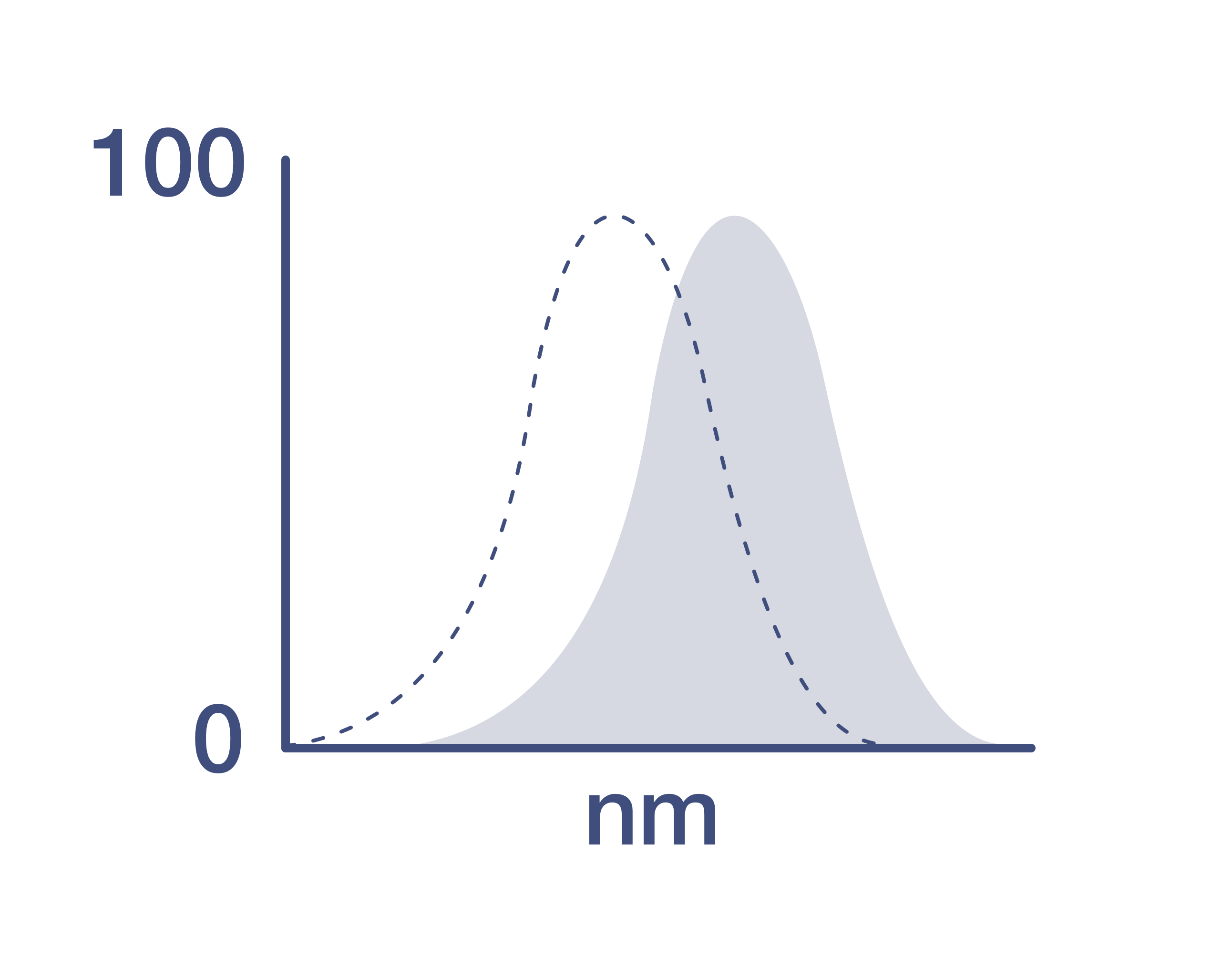
Phospho-AKT1 (Ser473) Antibody (12-9715-42) in Flow
Normal human peripheral blood cells were unstimulated (orange histogram) or stimulated with Lipopolysaccharide (LPS) Solution (500X) (Product # 00-4976-03) (purple histogram), then intracellularly stained with Anti-Human/Mouse phospho-AKT (S473) PE using the Intracellular Fixation and Permeabilization Buffer Set (Product # 88-8824-00) and protocol. Cell in the lymphocyte (left) and monocyte (right) gates we... View More


 Antibody (12-9715-42) in Flow")
 Antibody (12-9715-42) in Flow")
 Antibody (12-9715-42) in Flow")
 Antibody (12-9715-42) in Flow")
 Antibody (12-9715-42) in Flow")
 Antibody (12-9715-42) in Flow")
 Antibody (12-9715-42) in Flow")
 Antibody (12-9715-42) in Flow")
 Antibody (12-9715-42) in Flow")
 Antibody (12-9715-42) in Flow")
 Antibody (12-9715-42)")
Product Details
12-9715-42
Applications
Tested Dilution
Publications
Product Specifications
Species Reactivity
Human,
Mouse
Published species
Human,
Mouse
Host/Isotype
Mouse
/ IgG2a, kappa
Recommended Isotype Control
Class
Monoclonal
Type
Antibody
Clone
SDRNR
Conjugate
Excitation/Emission Max
565/576 nm
View spectra
Form
Liquid
Concentration
5 µL/Test
Purification
Affinity chromatography
Storage buffer
PBS, pH 7.2, with 0.2% BSA
Contains
0.09% sodium azide
Storage conditions
4°C, store in dark, DO NOT FREEZE!
Shipping conditions
Ambient (domestic); Wet ice (international)
RRID
AB_2637101
Product Specific Information
Description: This SDRNR monoclonal antibody recognizes human and mouse AKT (also known as Protein Kinase B (PKB)) when phosphorylated on S473. AKT is a serine/threonine protein kinase that plays a key role in multiple cellular processes including metabolism, proliferation, apoptosis/survival, and migration. There are three homologous isoforms of AKT: AKT1, AKT2, and AKT3. AKT is activated by binding of its pleckstrin homology (PH) domain to membrane phospholipids and by phosphorylation. Phosphorylation of AKT at T308 by PDK1 and at S473 is required for full activation of this kinase. AKT promotes cell survival by inhibiting apoptosis via phosphorylation and inactivation of several targets including Bad, Foxo1, c-Raf, and caspase-9. Deregulation of AKT has been implicated as a major contributing factor in many types of cancer. AKT is negatively regulated by the phosphatase PTEN as well as by the chemical inhibitor LY294002. Specificity of this SDRNR clone was determined by ELISA, flow cytometry, and western blotting.
Applications Reported:This SDRNR antibody has been reported for use in intracellular staining followed by flow cytometric analysis.
Applications Tested: This SDRNR antibody has been pre-titrated and tested by intracellular staining followed by flow cytometric analysis of normal human peripheral blood cells. This can be used at 5 µL (0.06 µg) per test. A test is defined as the amount (µg) of antibody that will stain a cell sample in a final volume of 100 µL. Cell number should be determined empirically but can range from 10^5 to 10^8 cells/test.
Staining Protocol: All protocols work well for this monoclonal antibody. Use of Protocol A: Two-step protocol: intracellular (cytoplasmic) proteins allows for the greatest flexibility for detection of surface and intracellular (cytoplasmic) proteins. Use of Protocol B: One-step protocol: intracellular (nuclear) proteins is recommended for staining of transcription factors in conjunction with surface and phosphorylated intracellular (cytoplasmic) proteins. Protocol C: Two-step protocol: Fixation/Methanol allows for the greatest discrimination of phospho-specific signaling between unstimulated and stimulated samples, but with limitations on the ability to stain specific surface proteins (refer to "Clone Performance Following Fixation/Permeabilization" located in the BestProtocols Section under the Resources tab online). All Protocols can be found in the Flow Cytometry Protocols: "Staining Intracellular Antigens for Flow Cytometry Protocol" located in the BestProtocols® Section under the Resources tab online.
Excitation: 488-561 nm; Emission: 578 nm; Laser: Blue Laser, Green Laser, Yellow-Green Laser.
Filtration: 0.2 µm post-manufacturing filtered.
Target Information
AKT1 (PKB alpha) is a serine/threonine kinase that regulates cell survival. The activated enzyme inhibits apoptosis and stimulates cell cycle progression by phosphorylating numerous targets in various cell types, including cancer cells. This protein kinase is activated by insulin, PI3K, IGF1 and various other growth and survival factors. Akt promotes cell survival by inhibiting apoptosis through phosphorylation and inactivation of several targets, including forkhead transcription factors, and caspase-9. The AKT pathway is a major target for cancer drug discovery.
For Research Use Only. Not for use in diagnostic procedures. Not for resale without express authorization.
How to use the Panel Builder
Watch the video to learn how to use the Invitrogen Flow Cytometry Panel Builder to build your next flow cytometry panel in 5 easy steps.
Bioinformatics
Protein Aliases: AKT1 kinase; AKT1m; Akt1m protein; C-AKT; CAKT; PKB; PKB alpha; PKB beta; PKB gamma; PKBG; Protein kinase B; Protein kinase B alpha; protein kinase B-alpha; Proto-oncogene c-Akt; rac protein kinase alpha; RAC-alpha serine/threonine-protein kinase; RAC-PK-alpha; RAC-PK-beta; RAC-PK-gamma; related to A and C kinases; serine-threonine protein kinase; Thymoma viral proto-oncogene; v-akt murine thymoma viral oncogene homolog 1; v-akt murine thymoma viral oncogene-like protein 1
Gene Aliases: AKT; AKT1; CWS6; PKB; PKB-ALPHA; PKB/Akt; PKBalpha; PRKBA; RAC; RAC-ALPHA
UniProt ID: (Human) P31749, (Mouse) P31750
Entrez Gene ID: (Human) 207, (Mouse) 11651
protein kinase activity
protein serine/threonine kinase activity
protein serine/threonine/tyrosine kinase activity
protein kinase C binding
protein binding
ATP binding
phosphatidylinositol-3,4,5-trisphosphate binding
kinase activity
enzyme binding
nitric-oxide synthase regulator activity
GTPase activating protein binding
identical protein binding
phosphatidylinositol-3,4-bisphosphate binding
protein phosphatase 2A binding
14-3-3 protein binding
nucleotide binding
transferase activity
transferase activity, transferring phosphorus-containing groups
protein kinase binding
protein import into nucleus, translocation
osteoblast differentiation
maternal placenta development
positive regulation of protein phosphorylation
positive regulation of endothelial cell proliferation
carbohydrate metabolic process
glycogen metabolic process
glycogen biosynthetic process
regulation of glycogen biosynthetic process
glucose metabolic process
translation
regulation of translation
protein phosphorylation
negative regulation of protein kinase activity
transport
apoptotic process
activation-induced cell death of T cells
inflammatory response
cytoskeleton organization
signal transduction
multicellular organism development
germ cell development
nervous system development
aging
insulin receptor signaling pathway
apoptotic mitochondrial changes
carbohydrate transport
response to hormone
negative regulation of autophagy
negative regulation of plasma membrane long-chain fatty acid transport
positive regulation of fibroblast migration
positive regulation of sodium ion transport
positive regulation of glucose metabolic process
negative regulation of endopeptidase activity
regulation of neuron projection development
glucose transport
phosphorylation
protein ubiquitination
peptidyl-serine phosphorylation
cell projection organization
protein catabolic process
positive regulation of cell growth
regulation of cell migration
regulation of myelination
positive regulation of cyclin-dependent protein serine/threonine kinase activity involved in G1/S transition of mitotic cell cycle
lipopolysaccharide-mediated signaling pathway
negative regulation of fatty acid beta-oxidation
response to food
positive regulation of cellular protein metabolic process
peripheral nervous system myelin maintenance
positive regulation of proteasomal ubiquitin-dependent protein catabolic process
cellular response to insulin stimulus
regulation of protein localization
positive regulation of peptidyl-serine phosphorylation
response to fluid shear stress
intracellular signal transduction
cellular response to vascular endothelial growth factor stimulus
cellular response to decreased oxygen levels
glucose homeostasis
anagen
positive regulation of apoptotic process
negative regulation of apoptotic process
negative regulation of cysteine-type endopeptidase activity involved in apoptotic process
protein kinase B signaling
positive regulation of blood vessel endothelial cell migration
positive regulation of nitric oxide biosynthetic process
positive regulation of fat cell differentiation
positive regulation of glycogen biosynthetic process
negative regulation of cell size
negative regulation of proteolysis
positive regulation of vasoconstriction
positive regulation of transcription from RNA polymerase II promoter
positive regulation of glucose import
negative regulation of JNK cascade
positive regulation of lipid biosynthetic process
insulin-like growth factor receptor signaling pathway
positive regulation of nitric-oxide synthase activity
positive regulation of sequence-specific DNA binding transcription factor activity
striated muscle cell differentiation
glycogen cell differentiation involved in embryonic placenta development
labyrinthine layer blood vessel development
response to UV-A
cellular response to mechanical stimulus
cellular response to growth factor stimulus
cellular response to epidermal growth factor stimulus
cellular response to prostaglandin E stimulus
cellular response to organic cyclic compound
cellular response to hypoxia
positive regulation of establishment of protein localization to plasma membrane
negative regulation of release of cytochrome c from mitochondria
cellular response to granulocyte macrophage colony-stimulating factor stimulus
execution phase of apoptosis
cellular response to peptide
cellular protein modification process
nitric oxide biosynthetic process
cellular response to DNA damage stimulus
response to oxidative stress
G-protein coupled receptor signaling pathway
cell proliferation
response to heat
negative regulation of gene expression
regulation of phosphatidylinositol 3-kinase signaling
peptidyl-threonine phosphorylation
spinal cord development
cell differentiation
platelet activation
hyaluronan metabolic process
endocrine pancreas development
T cell costimulation
positive regulation of endodeoxyribonuclease activity
ERBB2 signaling pathway
regulation of mRNA stability
positive regulation of epidermal growth factor receptor signaling pathway
protein autophosphorylation
phosphatidylinositol-mediated signaling
regulation of nitric-oxide synthase activity
response to growth hormone
mammary gland epithelial cell differentiation
establishment of protein localization to mitochondrion
maintenance of protein location in mitochondrion
chemical synaptic transmission, postsynaptic
negative regulation of protein kinase activity by protein phosphorylation
positive regulation of protein localization to nucleus
negative regulation of neuron death
regulation of signal transduction by p53 class mediator
regulation of cell cycle checkpoint
negative regulation of oxidative stress-induced intrinsic apoptotic signaling pathway
cellular response to nerve growth factor stimulus
response to insulin-like growth factor stimulus
negative regulation of extrinsic apoptotic signaling pathway in absence of ligand

Performance Guarantee
If an Invitrogen™ antibody doesn't perform as described on our website or datasheet,we'll replace the product at no cost to you, or provide you with a credit for a future purchase.*
Learn more
We're here to help
Get expert recommendations for common problems or connect directly with an on staff expert for technical assistance related to applications, equipment and general product use.
Contact tech support