Biomedical research leverages volume electron microscopy techniques
Volume electron microscopy allows biomedical researchers to study the 3D structure of cells, tissue and small organisms at nanometer resolution.
Because every organism has this type of multi-scale, 3D complexity, volume electron microscopy is relevant to understanding the secrets of human life and disease. It bridges the gap between light microscopy and cellular cryo-electron tomography and is extremely impactful for furthering 3D cell biology research.
It was also named one of Nature’s Seven Technologies to Watch in 2023.
The expanding community requires more dedicated, multipurpose and customizable life science solutions to help maximize instrument run times, ensure predictable and reproducible results, and continue to advance this method sure to create scientific breakthroughs yet-to-be-imagined.
Hydra Bio Plasma-FIB: A new dedicated solution for life sciences workflows
We’re excited to introduce our new Thermo Scientific Hydra Bio Plasma-Focused Ion Beam (Plasma-FIB) designed for cell biologists seeking simplified workflows while undertaking volume electron microscopy for cryo or resin-embedded samples.
Building off the revered Thermo Scientific Helios Hydra DualBeam platform, the Hydra Bio Plasma-FIB joins the company’s expanding cryo-FIB portfolio, including a suite of tailored solutions for a variety of life science needs, and is designed to be a bridge from cryo-EM to room temperature analysis or vice versa.
Register to attend a webinar about the new Hydra Bio Plasma-FIB on October 18 >>
From tissues to proteins, this versatile, multi-application instrument supports volume electron microscopy at cryogenic and room temperatures, as well as cellular cryo-electron tomography.
 |
“My lab is using cryo tomography to study the architecture of sarcomeres, the smallest subunits of striated muscle. Previously, we used isolated myofibrils to accomplish our goal. Now we are using the Hydra and cryo-lift out to be able to study muscle structures directly in the native muscle fibres.” |
|---|
Dr. Stefan Raunser, Director, Structural Biochemistry, Max Planck Institute of Molecular Physiology. Image courtesy of North Rhine-Westphalian Academy of Sciences and Arts / Bettina Engel-Albustin 2022.
The Hydra Bio Plasma-FIB addresses multiple workflows, including high-resolution serial PFIB imaging, including large area serial PFIB imaging using Thermo Fisher’s unique Spin Mill Bio Method, array tomography, correlative light and electron microscopy, and high-throughput lamella preparation for cryo-electron tomography.

Spin Mill Bio Method: A Case Study
The Spin Mill Bio Method used on the Hydra Bio Plasma-FIB allows researchers to uncover large sample areas and visualize regions of interest by providing large-area planar milling up to 1 mm in diameter, with a geometry like microtome-based serial block face imaging but at a slice thickness as small as 5 nm. Users can prepare clean, smooth surfaces used to localize regions of interest and subsequently image them in 2D or 3D.
In this example, by Watanabe et al, researchers used this method to study resin embedded mouse hippocampal synapses, high pressure frozen at different time points, and used tagging strategies to visualize different receptors and their relationship with synaptic vesicles.
For each sample, they milled a 638 µm wide area view of the tissue and identified sparsely distributed synapses within the sample. The tagging of the receptors at these locations with gold enabled fast and easy end pointing and stacking of images.
They then zoomed in on these regions of interest at high resolution, delivering images that they could use to measure the critical dimensions of the receptors in which they were interested.
Being able to compare different samples, frozen at specific time points in a fast and time efficient manner, enabled a deeper understanding of synaptic function. The Hydra Bio Plasma-FIB has both the speed and the resolution needed to understand physiological processes at the nanometer level.
Advancing cell biology research through technological innovation
The further systematic buildup of a volume electron microscopy community will continue to fuel technological developments as well as provide access for increasingly large groups of bioscience researchers to benefit from these exciting and valuable techniques.
 |
“We are increasingly being challenged to understand how to improve crop resilience to environmental stress and disease. The remarkable capabilities of our new Hydra will allow us the unprecedented ability to reconstruct entire plant cells and tissue with exquisite detail. This technology will allow us to “freeze” organisms in time and space and build intricate 3D models that will help us solve our critical food security challenges.” |
|---|
Dr. Kirk J. Czymmek, Principal Investigator, Director Advanced Bioimaging Laboratory, Donald Danforth Plant Science Center
At Thermo Fisher, we’re focused on these technological developments so scientists can focus on what’s most important—their research and the positive impact it continues to have on our understanding of life and disease.
With the new Hydra Bio Plasma-FIB, we’re proud to offer cell biologists access to a life-science-dedicated cryo-FIB system that is easier to use and helps simplify room temperature and cryogenic workflows—similar to what we’ve developed for cryo-EM single particle analysis needs over the last decade.
Learn more about the Thermo Scientific Hydra Bio Plasma-FIB >>
Register to attend ‘Cryo-tomography to Volume EM: Explore with Hydra Bio Plasma-FIB’ on Oct. 18 >>


Postmortem brain tissue imaging sheds light on cognitive function in life
Nanoscale brain imaging with electron microscopy Communicati...
Read More
Decoding tauopathies: how cutting-edge cryo-EM is unraveling tau protein and Alzheimer’s disease
Tauopathy and neurodegeneration Neurodegenerative diseases a...
Read More
Virus microscopy unravels chikungunya virus structure
Global impacts of the chikungunya virus Chikungunya is a vir...
Read More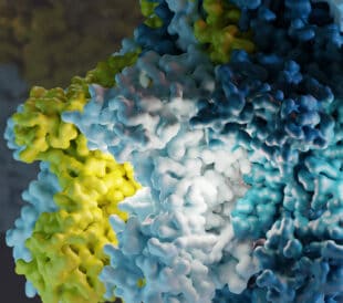
What is cryo-EM?
Cryo-EM: cryo electron microscopy Cryo-electron microscopy (...
Read More
Leave a Reply