There are many different apoptosis assays for flow cytometry because cell death cascades are complex and dynamic, underscoring the importance of a multi-parametric approach to the assessment of apoptosis. Since no single parameter defines programmed cell death, a combination of techniques is recommended for flow cytometry apoptosis detection.

Flow cytometry apoptosis detection assays
We offer a wide variety of apoptosis assays for flow cytometry that detect changes in the plasma and mitochondrial membranes, caspase activity, nuclear condensation, and fragmentation as possible indicators of apoptosis. Mix and match our standalone reagents or choose from our selection of multi-parametric apoptosis assay kits that are designed to selectively differentiate apoptotic cells from living and necrotic cells in a single cell population.

Annexin V staining is used to detect the translocation of phosphatidylserine to the outer leaflet of the plasma membrane, a hallmark of apoptosis.
- Available as stand-alone reagents or easy-to-use kits
- Alternative reagents if the required calcium buffer or the trypsinization of adherent cells doesn’t fit your workflow

The activation of caspase enzymes is a distinctive feature of apoptosis.
- Assays for Caspase 3/7, 8 and poly caspase activity
- Multiple detection options: fluorogenic substrate or fluorescent inhibitors
- Multi-parameter kits for identifying live, dead, and apoptotic cells

Mitochondrial dysfunction is a feature of apoptosis. A wide variety of reagents are available to detect:
- Membrane potential by dynamic changes or end point assays
- Increased superoxide production
- Influx of calcium influx into mitochondria

Apoptosis is characterized by changes in nuclear morphology, including DNA fragmentation, chromatin condensation, and DNA strand breaks.
- TUNEL assays
- DNA stains
Selection Guide: search apoptosis assays for flow cytometry
Select flow cytometry apoptosis detection reagents based on the laser excitation source and common emission filters.
UV laser (~350 nm) reagents
| Emission filter | 379/28 nm | 440/40 nm |
| Annexin V conjugates | ||
| Membrane permeability & chromatin condensation |
Violet laser (405 nm) reagents
| Emission filter | 450/40 nm | 525/50 nm | 610/20 nm |
| Annexin V conjugates | |||
| Annexin V alternative (when the use of calcium containing buffers isn’t an option) | |||
| Membrane permeability & chromatin condensation |
Blue laser (488 nm) reagents
| Emission filter | 530/30 nm | 574/26 nm | 695/40 nm | 780/60 nm |
| Annexin V conjugates | ||||
| Caspase activity | ||||
| Membrane permeability & chromatin condensation | ||||
| Dynamic changes in mitochondrial membrane potential | ||||
| End point assays for mitochondrial membrane potential | ||||
| Mitochondrial transition pore | ||||
| TUNEL assays |
Green laser (532 nm) reagents
| Emission filter | 585/16 nm | 620/15 nm |
| Annexin V conjugates | ||
| End point assays for mitochondrial membrane potential | ||
| Mitochondrial superoxide production | ||
| Mitochondrial calcium influx |
Yellow laser (561 nm) reagents
| Emission filter | 585/16 nm | 620/15 nm |
| Annexin V conjugates | ||
| Dynamic changes in mitochondrial membrane potential | ||
| End point assays for mitochondrial membrane potential |
Red laser (637 nm) reagents
| Emission filter | 670/14 nm |
| Annexin V conjugates | |
| Membrane permeability & chromatin condensation | |
| End point assays for mitochondrial membrane potential |
Learn more about
Resources
Fluorophore and reagent selection guide for flow cytometry
Download Flow Cytometry Protocols Handbook
Spectral Flow Cytometry Fundamentals
Invitrogen eBioscience Resources—Selection guides, Best Protocols, product performance and more.
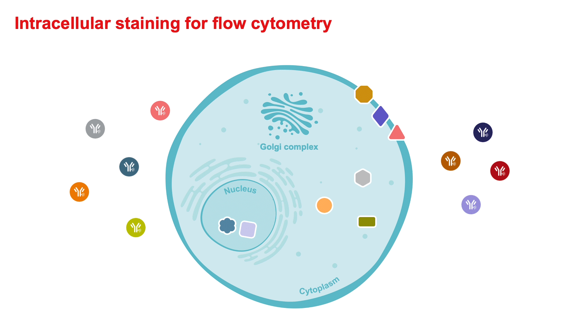
Intracellular Staining for Flow Cytometry How-To Video—for detecting cytokines and intranuclear markers.
Flow Cytometry Learning Center—Access flow cytometry educational resources for better experiment planning and execution.
Flow Cytometry Panel Builder—Design your flow cytometry panel with this online tool for a simplified, customizable experience to fit your needs.
5 Steps Resources
Support
Flow Cytometry Support Center—Find technical support recommendations for your flow cytometry workflows, including tips for experimental setup and in-depth troubleshooting help.
Flow Cytometry Panel Design Support—Work with one of our technical sales specialists to discuss your experimental needs and guide you through the process.
Not for resale. Super Bright Polymer Dyes are sold under license from Becton, Dickinson and Company.
For Research Use Only. Not for use in diagnostic procedures.